- About
- What We Do
- Initiatives
- Research
- News
- Get In Touch
Back to Top Nav
Back to Top Nav
Back to Top Nav
The new instrument offers ultra-high-res images and 3D reconstructions.
In the basement of Remsen Medical Sciences Building, one of the hubs of Dartmouth's research enterprise has quietly been ramping up, stronger than ever.
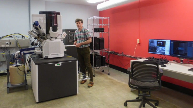
When the pandemic closed campus this spring, the Electron Microscope Facility had just finished installing a new, next-generation scanning electron microscope (SEM)—a Thermo Fisher Scientific Helios 5 CX DualBeam capable of producing ultra-high-resolution imaging and three-dimensional reconstructions, allowing scientists to cut using a focused ion beam (FIB) and see under the surface of the materials they study.
"Instrumentation like this is essential if Dartmouth students and faculty are to drive learning and discovery at an internationally competitive level," says Dean Madden, vice provost for institutional research.
"Despite the pandemic, the new Helios microscope was installed in record time, thanks to a team effort led by the facility director, Dr. Maxime Guinel," Madden says. "The instrument is already heavily used, providing state-of-the-art results for a wide range of projects in chemistry, material sciences, and physics, and will soon expand to include new biological applications."
The Helios microscope replaced an older Scios model that was irreparably damaged during heavy rainstorms last year that flooded parts of Remsen. Guinel, who has overseen the facility since 2018, says the replacement is a substantial improvement.
"The fifth-generation of the Helios family was just commercialized when we were looking for a replacement, and we were able to get the latest model at a reduced cost," Guinel says. "It gives us a better resolution and enhanced capabilities to carry on research projects."
In addition to the Helios, the facility offers easy access to a range of other microscopes and imaging instruments, as well as training and support for students and faculty from across the institution who want to use the microscopes in their research (socially distanced access to the facility is available by appointment). The facility also has a lower-resolution scanning electron microscope housed at Thayer School of Engineering.
"The acquisition of the Helios scanning electron microscope has greatly enhanced the research capabilities at Dartmouth in terms of imaging resolution, sensitivity, access to state-of-the-art electron microscope technologies, and versatility in combining several modes of analysis," says Radu Stan, an associate professor of biochemistry and cell biology and of pathology and laboratory medicine at the Geisel School of Medicine.
Crucially, the microscope allows researchers to do what is known as volume or FIB-SEM microscopy experiments that combine a focused ion beam, scanning electron microscopy, and 3D reconstructions—a method "which was not previously able to be done at Dartmouth," Stan says. Stan's lab studies the role of specific endothelial subcellular organelles in mammalian vascular permeability.
"The ability to do FIB-SEM imaging is an exciting development for life sciences researchers, allowing high resolution imaging and visualization of subcellular structures in tissues and cells in 3D," Stan says. "The availability of this instrument at Dartmouth gives our researchers a clear competitive advantage on the type of scientific questions they are able to ask, which will translate in increased depth of inquiry, more effective science and possibly a greater success in securing external research funding."
Katherine Mirica, an assistant professor of chemistry who studies the relationship between the structural properties of materials and their functions, calls the new microscope "absolutely crucial for the characterization of nanostructured materials that my group synthesizes"—materials with potential applications in chemical sensing, personal protective equipment, and energy conversion and storage.
"The most exciting thing about this microscope is the ability to obtain high resolution information about the structure of nanomaterials with co-localized elemental mapping," Mirica says. "Scanning electron microscopy is an important technique for promoting scientific discovery in materials chemistry and nanoscience aimed at addressing global challenges in healthcare, environmental stewardship, and energy-related research. Having this instrument accessible across the institution allows for collaborations across multiple disciplines in tackling these major challenges that the world faces today."
Next year, Guinel and the Electron Microscope Facility will move to their permanent home in the new technology hub being built on the West End of campus that will be shared by Thayer School of Engineering, the Department of Computer Science, and the Magnuson Center for Entrepreneurship.
"When we move to the new facility taking shape in the West End, the Helios will anchor our suite of instruments," Madden says.
Hannah Silverstein can be reached at hannah.silverstein@dartmouth.edu.